A link between a specific brain protein and susceptibility to neurodegeneration in people with epilepsy has been uncovered by Florida State University College of Medicine researchers. This work has the potential to expand neurology frameworks related to epilepsy and neurodegenerative disease progression, potentially improving clinical outcomes.
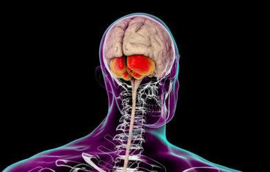
Temporal lobe epilepsy (TLE) is a form of epilepsy most commonly seen in adults, and often these patients do not see relief with medication. The research team took a novel approach to studying small amounts of tissue from more remote areas in the brain. They focused on glutamate and a receptor of it called N-methyl-D-aspartate (NMDA). Glutamate is an important chemical that plays a key role in memory and learning, but it has to be in the right amount in order to function most optimally.
The team discovered that although two proteins commonly associated with NMDA, GluN1 and GluN2, were evenly distributed in the hippocampal region (the learning and memory center of the brain), a third one called GluN3 was distributed on a gradient. Neuronal loss in this region of the brain is almost a dead giveaway that TLE is progressing. “The relationship between GluN3 and cell loss was not known until this research,” says Sanjay Kumar, professor of Biomedical Sciences, in a statement. “This advance in cellular biology is an important step for developing therapies to help patients.”
Since GluN3 makes neurons more likely to suffer from calcium-induced cellular damage, the finding helps researchers focus on identifying precisely where neurons are dying in a specific area, especially those hard to reach. To expand on this work, Kumar has applied to patent the technique, known as area-specific tissue analysis (ASTA), that he developed himself. ASTA’s accuracy has provided an improved way of testing for both the presence and amount of specific proteins linked to TLE. “This research shows how area-specific tissue analysis can be a useful tool,” he says. “I’m excited to explore what further research with this technique can uncover.”
The team is very proud of their work, and hope that it inspires other neurology researchers to conduct more related studies that can promote more effective therapeutic interventions. ASTA is a useful tool that can aid this, allowing researchers to examine parts of the brain that are not easily explored. The gradient distribution of GluN3 will allow scientists to access more pertinent samples of tissue that have a greater impact on neurodegeneration, and not just the ones that are able to be reached the easiest.
The study is published in the journal Neurophysiology.


-392x250.jpg)