In a new, and rare study of direct EEG recordings in children and adolescents, a Northwestern Medicine scientist and colleagues from Wayne State University have discovered that as brains mature, the precise ways by which two key memory regions in the brain communicate with each other, forming lasting memories. The quality of the study was made possible using intracranial EEG – new technology in which these scientists were pioneers.
Previous research using high-resolution external EEG data from children’s brains left gaps in understanding of how the developing brain forms memories.
The scientists found a link between how the brains of people ages 5 to 21 were developing and how well they were able to form memories throughout that 16-year period. For example, younger children weren’t able to form as many memories as some adolescents.
“Our study helps us actually explain how memory develops, not just that it develops,” says Lisa Johnson, assistant professor of medical social sciences and pediatrics at Northwestern’s medical school, in a statement. “By understanding how something comes to be — memory, in this instance — it gives us windows into why it eventually falls apart. Human memory develops throughout childhood, peaks in your 20s and, for most people, declines with age, even in those who don’t develop dementia.”
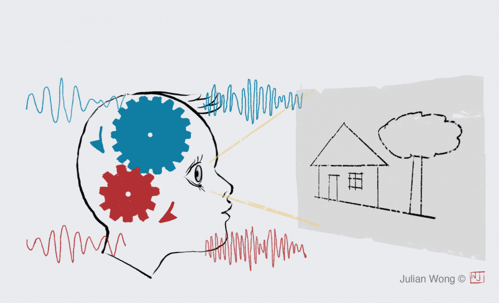
The study focused on communication between two regions of the brain that play a key role in supporting memory formation: the medial temporal lobe (MTL) and prefrontal cortex (PFC). To learn how these regions communicate, the scientists analyzed two brain signals — a slowly oscillating brain waves and a faster oscillating brain wave. These enable communication between regions. The rhythms corresponded to both success in forming memories and quality of performance.
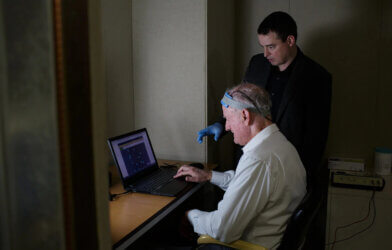
Participants in the study were undergoing brain surgery for another reason (usually epilepsy), creating this rare opportunity to collect data from electrodes placed directly on brain tissue.
The study is published in the journal Current Biology.