The unending intricacies of how the human brain works continues to remain a mystery. To understand its vast complexity, scientists have long studied the cellular makeup of the brain, along with the communications that take place within it. Researchers at the Baylor College of Medicine are making progress, revealing insights into how the brain functions through a model called Spatial Transcriptomics cell-types Assignment using Neural Networks, or STANN.
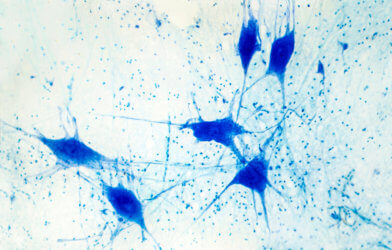
Currently, scientists are able to locate individual cells within the brain and determine the function and product of each cell. The brain, however, is made up of several millions to hundreds of billions of cells. The challenge arises in collecting the data in order to gather a complete understanding of how the brain works.
STANN leads to ‘eureka moment’
Samee and his colleagues worked to create a clear and comprehensible model of the brain as well as the functions of its tissues. The team applied STANN, along with other methods and datasets, and began to see more clear patterns and functions of cells within the brain.
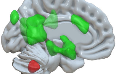
The brain has evolved to consist of distinct layers, referred to as morphological layers. By using their findings, Samee and his colleagues were able to study samples from each layer in order to understand the general makeup. Overall, the team found that the makeup remained consistent within each layer. “However, the percentages may change from one layer to another,” Samee explains in a media release. “Layer by layer, our model provided the precise location of different cell types, whether they communicated with each other and by which means,” Samee said. “This was a ‘eureka moment’ for us.”
The team was also able to make groundbreaking discoveries regarding the locations and interactions of cells across each morphological layer of the brain. In one area of the layer, for instance, populations of cells and neurons may be grouped together but found separate in another section of the same layer. Samee says that there is communication between cell types within a layer, revealing that gene networks change based on their location.
‘Detailed view of brain organization not described before’
These studies have helped the researchers to better understand that each layer of the brain is composed of specific cell types, though the grouping of these types changes throughout the layer. It is hypothesized, then, that each cell type has a subtype, which performs specific functions based on its location. “This new detailed view of brain organization at the single-cell and functional levels had not been described before,” notes Samee.
“The neural network model approach we have developed in this work presents an ‘instruction manual’ for other researchers to use to study other areas of the brain or other organs, such as the heart, the main interest of my lab,” says co-author Dr. James Martin, vice chairman and professor of molecular physiology and biophysics at the medical school, and director of the Center for Organ Repair and Renewal.
Article written by Rhonda Errabelli