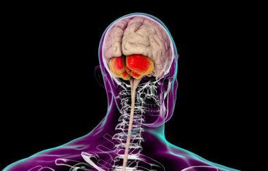
Advances in medical technology used for imaging — such as X-rays, CT scans, magnetic resonance imaging — are so great that the field can seem more like science fiction than tools we use every day. Now, using similarly-impressive special techniques, Yale researchers have confirmed that the destruction of neuron-to-neuron synapses within the brain are the source of the cognitive deficits characteristic of Alzheimer’s disease.
The incidence of Alzheimer’s disease (the number of people per 100,000 who newly develop the disease per year) seems to be declining. The lesser rate has been attributed to improvements in Alzheimer’s risk factors, such as hypertension. Even with this potentially lower incidence, however, the number of people with Alzheimer’s disease is expected to continuously increase, because the population of adults aged 65 and older is increasing. Most of the baby-boom generation (Americans born between 1946 and 1964) have already reached age 65; the oldest members of the baby-boom generation turned aged 75 in 2021.
Lifetime risk is the probability that someone of a given age who does not have a condition will develop the condition during that person’s remaining life span. Data from another study were used to estimate lifetime risks of Alzheimer’s dementia by age and sex. The study shows that the estimated lifetime risk for Alzheimer’s dementia at age 45 was approximately 1 in 5 for women and 1 in 10 for men. The risks for both sexes were slightly higher at age 65.
In the past, scientists assumed that the loss of connections between brain cells caused Alzheimer’s-type symptoms, but actual evidence of the synaptic loss had been limited to autopsy. A new technology was needed to evaluate disease in living patients. Yale scientists developed a positron emission tomography (PET) scanning technology, which allows researchers to observe the loss of synapses in living patients with even mild Alzheimer’s disease.
The new glycoprotein 2A (SV2A) PET imaging scan allows scientists to measure metabolic activity at the brain synapses of people diagnosed with mild to moderate Alzheimer’s disease. Then they measured each person’s cognitive performance in verbal memory, language skills, executive function, processing speed, and visual-spatial ability.
They report that the loss of synapses or connections between brain cells was strongly associated with poor performance on cognitive tests. Results also reveal that synaptic loss was a stronger indicator of poor cognitive performance than was the loss of volume of the neurons in the brain.
Study authors say the loss of synapses in patients over time yields better understanding of cognitive decline in individuals. “The findings help us understand the neurobiology of the disease and can be an important new biomarker to test the efficacy of new Alzheimer’s drugs,” explains Adam Mecca, assistant professor of psychiatry at Yale, in a statement.
The research is published in the journal Alzheimer’s & Dementia: The Journal of the Alzheimer’s Association.


-392x250.jpg)